Biology
AI Foundation Model Maps Cancer Tissue: VirTues
VirTues, a new foundation model published in Nature, learns one shared representation of spatial proteomics data across marker panels and imaging platforms—and finds biomarkers that predict immunotherapy response.
One AI Model to Read Every Cancer Tissue Map#
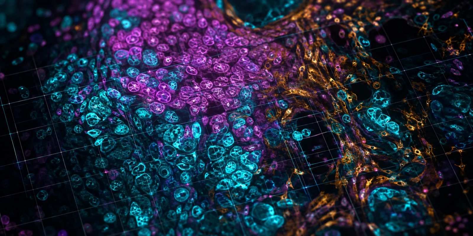
For fifteen years, pathologists and cancer biologists have been building ever more detailed pictures of what tumours actually look like on the inside: not just which cells are present, but how they are arranged, which immune cells sit next to which cancer cells, and what molecular signals pass between them. The tools that produce these pictures, known collectively as spatial proteomics, can now measure dozens to hundreds of proteins in a single slice of tissue while keeping every measurement pinned to its exact location. The result is a map of a tumour at near-single-cell resolution.
There is a catch that has quietly held the field back. Almost every lab builds its map differently. One group stains for 40 proteins; another picks a different 50; a third uses a separate imaging machine entirely. Each dataset ends up speaking its own dialect, and software written for one study usually cannot read another. A biomarker that looks promising in one cohort often falls apart when someone tries to reproduce it elsewhere.
On 5 August 2026, a team led by researchers at EPFL in Switzerland published a paper in Nature describing a system built to solve exactly this problem. It is called VirTues (short for Virtual Tissues), and it is a foundation model for spatial proteomics [1]. The claim is ambitious: a single trained model that can read tissue maps regardless of which proteins were measured or which machine took the image, and that turns fragmented, incompatible datasets into one searchable atlas.
Executive Summary#
VirTues is a general-purpose AI model trained on the largest open collection of spatial proteomics data assembled to date: 32 clinical cohorts, more than 5,100 patients, and 239 distinct protein markers across four imaging technologies [1]. Its central trick is to describe each protein marker not by an arbitrary channel number but by an embedding drawn from a protein language model, the same class of AI that powers modern structure prediction. That lets the model place different antibody panels into a shared representational space and interpret markers it never encountered during training. From one pretrained backbone, VirTues performs cell segmentation and typing, reconstructs unmeasured markers, annotates tissue neighbourhoods, retrieves similar patient cases, and discovers spatial biomarkers. In triple-negative breast cancer, its learned signatures predicted response to anti-PD-L1 chemo-immunotherapy and separated patients by survival better than previously published biomarkers and current clinical staging. The work is open access, and the code is public.
What Happened?#
The paper, "The Virtual Tissues foundation model resolves spatial proteomics across scales," appeared as an open-access article in Nature on 5 August 2026 [1]. It is the peer-reviewed version of work the group first posted as a preprint in early 2025 [3].
VirTues is trained on multiplexed tissue images, chiefly from imaging mass cytometry, with additional data from the CODEX, Orion, and MIBI platforms. The core training resource comprises 15 imaging mass cytometry datasets covering 3,102 patients and 146 markers across eight organ sites; the full corpus extends to 32 cohorts and 239 markers [1]. Rather than treating each image channel as an anonymous layer, VirTues represents every marker through embeddings from ESM-2, a protein language model [5]. Because those embeddings encode something about each protein's sequence and biology, two panels that share few literal markers can still be compared through the proteins' underlying relationships.
The architecture is a transformer redesigned for this data. Standard vision transformers scale poorly when an image has dozens of channels, because their computation grows quadratically with both space and channel count. VirTues splits attention into two components: one that reasons across markers and one that reasons across space, so it can handle high-plex images efficiently [1]. It is trained by masked autoencoding: hide parts of the tissue, then learn to reconstruct them. From that single objective, the model produces representations at four nested scales (protein, cell, niche, and tissue), each usable for a different downstream task.
Background: The Vocabulary You Need#
A few concepts make the rest of this clearer.
Spatial proteomics measures many proteins at once inside intact tissue while preserving where each protein sits. Compared with older methods that grind tissue into a slurry and lose all geometry, it reveals which cell types cluster together and how the tumour microenvironment (the ecosystem of cancer, immune, and structural cells) is organised.
A foundation model is a large model trained once on broad data by self-supervision, then reused across many tasks without starting from scratch each time. Large language models are the familiar example; the same recipe has since reshaped protein structure prediction and single-cell biology [1].
A protein language model treats a protein's amino-acid sequence like text and learns statistical patterns from millions of sequences. Systems such as ESM-2 use those patterns to predict structure and function [5]. VirTues borrows these embeddings to give each marker a meaningful identity instead of a bare label.
Zero-shot means the model handles something it was never explicitly trained on (here, a marker absent from its training panels) by reasoning from what it already knows.
Why This Matters#
The immediate payoff is interoperability. Spatial proteomics has generated a growing archive of richly annotated tumours, but that archive has behaved like a shelf of books in mutually unintelligible languages. A model that maps them into one space makes it possible, in principle, to pool cohorts, reuse annotations, and test whether a biomarker holds up across studies rather than within a single one [1].
The clinical thread is the more striking part. Using data from a published trial of triple-negative breast cancer, VirTues learned spatial signatures that predicted which patients responded to anti-PD-L1 chemo-immunotherapy, and it stratified disease-free survival in a separate, independent cohort [1]. These are not single-protein markers or simple cell counts. They are multicellular patterns (the arrangement and phenotype of many cell populations at once) which older approaches struggle to capture [4]. If such signatures generalize, they point toward richer, image-based tools for deciding who benefits from immunotherapy, a question where current predictors remain frustratingly unreliable.
More broadly, VirTues is a concrete step toward what its senior authors have called the "AI virtual cell": a computational representation of biological systems that can be queried, compared, and reused [2]. Spatial proteomics is one slice of that vision, but a telling one, because it links molecular detail to tissue-scale structure and, ultimately, to patient outcomes.
Critical Analysis#
The strengths are real: a large and genuinely heterogeneous training set, an architecture designed for the quirks of multiplexed imaging, open weights and open data, and results that hold up on tasks spanning segmentation to survival prediction. The protein-language-model tokenization is an elegant answer to the panel-incompatibility problem that has dogged the field.
The limits deserve equal attention. The model is developed and evaluated primarily on imaging mass cytometry; while the authors report strong cross-technology transfer, the bulk of the evidence still leans on one platform [1]. The headline clinical results come from retrospective analysis of existing trial and cohort data, not from prospective testing, so they show associations, not proven clinical utility. The independent-cohort validation is encouraging but narrow, focused on specific breast-cancer settings. And as with any foundation model, performance depends on the diversity of the training data; rare cancers, underrepresented populations, or unusual antibody panels may not be served as well as the common cases that dominate the corpus.
There are also open questions the paper does not settle. How stable are the discovered biomarkers across scanners, staining batches, and clinical sites in routine practice? How interpretable are the multicellular signatures to a pathologist who has to act on them? And how far does "technology-agnostic" really stretch once the imaging modality changes substantially? None of this diminishes the contribution; it marks the distance between a strong research result and a clinical tool. Realistically, prospective validation and regulatory review put any diagnostic use several years out.
Expert Perspective#
VirTues arrives in a lineage of biological foundation models. Protein language models such as ESM demonstrated that self-supervision could capture deep structure from sequence alone [5]. Single-cell models like scGPT extended the idea to expression data, and pathology foundation models brought it to whole-slide images. What distinguishes VirTues is the target: high-plex spatial proteomics across incompatible panels, a setting where earlier encoders assumed a fixed panel and simply could not relate markers they had not been trained on [1].
Competing efforts exist. Other groups have proposed foundation models for spatial or microscopy data, and the paper benchmarks against several, including recent spatial-proteomics models and masked-autoencoder baselines [1]. The differentiator VirTues offers is the marker-aware tokenization that makes heterogeneous panels comparable, combined with a multi-scale readout that serves both single-cell tasks and cohort-level discovery from the same backbone. Whether it becomes the field's default or one option among several will depend on how well independent labs reproduce its cross-cohort claims on their own data.
Key Takeaways#
- VirTues is a foundation model for spatial proteomics, published open access in Nature on 5 August 2026, trained on 32 cohorts, 5,100+ patients, and 239 markers across four imaging platforms [1].
- It represents each protein marker through protein-language-model embeddings, letting one model read different antibody panels and interpret markers it never saw during training [1][5].
- From a single pretrained backbone it handles cell segmentation and typing, marker reconstruction, niche annotation, case retrieval, and biomarker discovery [1].
- In triple-negative breast cancer, its spatial signatures predicted anti-PD-L1 chemo-immunotherapy response and stratified survival in an independent cohort, beating prior biomarkers and clinical staging [1][4].
- The clinical findings are retrospective and platform-weighted toward imaging mass cytometry; prospective validation is still needed before any diagnostic use.
Frequently Asked Questions#
What is spatial proteomics, in one sentence? It is a family of techniques that measures many proteins at once inside a tissue slice while recording exactly where each protein is, producing a molecular map of the tissue.
What makes VirTues a "foundation model" rather than just another algorithm? It is trained once, by self-supervision, on a large and varied dataset, and then reused across many tasks without retraining for each, the same pattern behind large language and protein models [1].
Why does using a protein language model matter here? It gives each marker a biologically meaningful identity based on the protein's sequence, so panels that share few literal markers can still be compared and the model can reason about proteins it never saw during training [1][5].
Does this mean AI can now diagnose cancer from tissue images? No. VirTues shows strong retrospective performance on research cohorts, but the clinical results are associations, not proven diagnostic utility. Prospective trials and regulatory review would be required before clinical use [1].
Is it available to other researchers? Yes. The paper is open access and the code is released publicly, alongside what the authors describe as the largest open collection of spatial proteomics data to date [1][6].
How is this different from AlphaFold-style models? AlphaFold-class systems predict the 3D structure of individual proteins. VirTues works one level up, reading how many proteins and cells are organised within whole tissues, and connecting that organisation to clinical outcomes [1].
What is the "AI virtual cell" this is said to advance? It is a longer-term goal of building computational models of biological systems that can be simulated and queried; spatial proteomics is one component, linking molecules to tissue structure [2].
A Note on Evidence#
This article summarizes a peer-reviewed paper and provides context, not medical, clinical, or investment advice. Where the original work reports retrospective or association-based findings, that distinction is preserved here. Claims about clinical utility remain hypotheses pending prospective validation.
References#
[1] Wenckstern, J., Jain, E., von Querfurth, B., Cheng, Y., et al. "The Virtual Tissues foundation model resolves spatial proteomics across scales." Nature (2026). Open access. https://doi.org/10.1038/s41586-026-10884-y
[2] Bunne, C. et al. "How to build the virtual cell with artificial intelligence: priorities and opportunities." Cell 187, 7045–7063 (2024). https://doi.org/10.1016/j.cell.2024.11.015
[3] Wenckstern, J. et al. "AI-powered virtual tissues from spatial proteomics for clinical diagnostics and biomedical discovery." Preprint (not peer reviewed), arXiv (2025). https://arxiv.org/abs/2501.06039
[4] Wang, X. Q. et al. "Spatial predictors of immunotherapy response in triple-negative breast cancer." Nature 621, 868–876 (2023). https://doi.org/10.1038/s41586-023-06498-3
[5] Lin, Z. et al. "Evolutionary-scale prediction of atomic-level protein structure with a language model." Science 379, 1123–1130 (2023). https://doi.org/10.1126/science.ade2574
[6] VirTues code repository, bunnelab/virtues. https://github.com/bunnelab/virtues