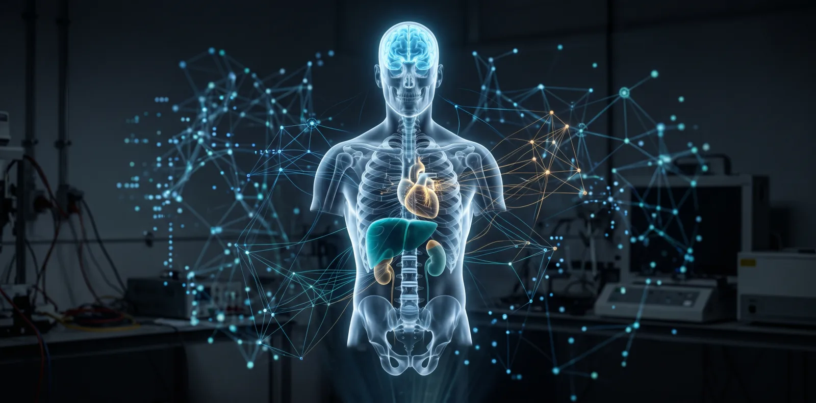
Two people turn 55 on the same day. One has lungs that look 62 and kidneys that look 48. The other is the reverse. Nothing on either birth certificate captures that, and until recently nothing in a clinic could either.
A study in Nature Medicine, published on 14 August 2026 by a team in Vienna trained computer vision models on 25,712 microscope slides of human tissue and found that organs keep separate calendars. The part that should make you sit up: a usable fraction of that organ-level signal leaks into the bloodstream, where an ordinary gene expression profile can pick it up.
What happened#
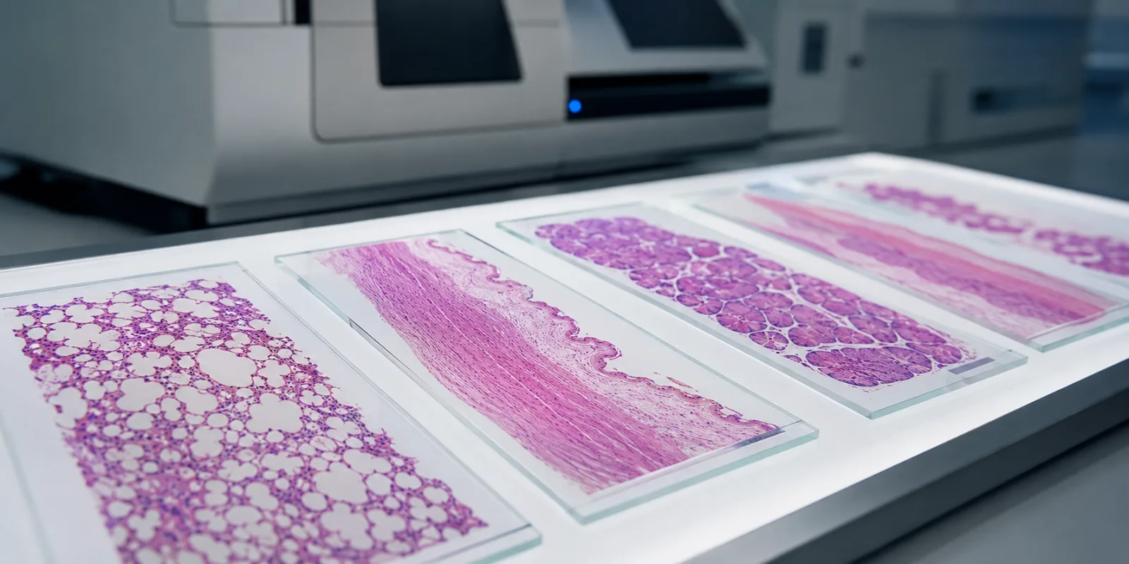
Researchers at the CeMM Research Center for Molecular Medicine in Vienna, working with the Ludwig Boltzmann Institute for Network Medicine, took the image archive of the Genotype-Tissue Expression project and asked a question nobody had asked at that scale. GTEx collected tissue from 983 deceased donors aged 20 to 70, covering 40 tissue types across 29 organs, all reviewed by pathologists. That is 25,712 whole-slide images, which the team broke into roughly 480 million small tiles and fed through vision models.
Age turned out to be the single strongest factor shaping how tissue looks, across all 40 tissue types, even though the models were never told to look for it. Building on that, the team trained per-tissue regression models they call tissue clocks. Average error was 4.88 years against known chronological age, with a coefficient of determination of 0.69.
Then they did the harder thing. They matched each donor's tissue-derived age gap to that same donor's blood gene expression and trained predictors that estimate organ-specific biological age from blood alone. Applied to 1,205 blood samples from nine independent public cohorts, those predictors picked out the right organ for the right disease: the brain in Alzheimer's disease, the gastrointestinal tract in Crohn's disease, the kidney, liver and heart in vasculitis.
What an ageing clock actually measures#
Two numbers describe how old you are. Chronological age counts birthdays. Biological age tries to describe the physical state of your cells and tissues, and the two come apart constantly. The difference between them is the age gap, and it is the quantity everyone in this field actually cares about.
The idea goes back to Steve Horvath's 2013 DNA methylation clock, which predicted age from chemical tags on DNA. Methylation is a molecular readout. This new work reads something different: architecture. When a pathologist looks at a tissue slide, they see structure, how cells are packed, how vessels branch, where scar tissue has formed. That structure changes with age, and a neural network can quantify the change far more finely than a human eye.
Two technical pieces made this possible. Pathology foundation models are large networks pretrained on millions of tissue images that produce a numerical fingerprint for any slide, without needing task-specific training. Vision-language models go further and let you ask an image how similar it is to a written phrase. The team used one to query 150 histological terms and found that atrophy, fibrosis and microvascular rarefaction, the thinning out of small blood vessels, rose with age across many organs, while markers of epithelial renewal fell.
Why this matters#
The clinical prize is a blood test that flags which organ is in trouble before symptoms appear. We are not there. But this is a credible route, and it comes with something the field has been short of: an interpretable account of what the model is looking at. Fibrosis and vessel loss are things a pathologist can point to on a slide, which makes the output arguable rather than oracular.
For researchers, the more useful contribution is the map. Lung, kidney, pancreas and adrenal gland showed accelerated ageing as early as the twenties and thirties. The uterus shifted sharply around menopause. Kidney failure tracked with accelerated ageing across several tissues, and the strongest signal was not in the kidney at all but in adipose tissue, pituitary, spleen and tibial nerve. Diabetes hit the pancreas hardest. These are hypotheses with tissue-level evidence attached.
One methods note for anyone building clocks: regularised linear models and ensembles generalised well to external cohorts, while neural network regressors did substantially worse. Bigger did not win.
Critical analysis#
Start with the strengths. The dataset is unusually good, the external validation is real, and the work is reproducible: the code and processing library are on GitHub. The authors benchmarked their fine-tuned model against 7 classical vision models and 18 pathology foundation models rather than declaring victory on one architecture.
Now the limits, which the authors state plainly.
The cohort is postmortem and cross-sectional, so every association is a correlation, and validation used people who already had a diagnosis. Whether an elevated organ age gap in blood appears before disease onset is untested. Until a prospective cohort with pre-diagnostic samples answers that, this describes illness rather than predicting it.
The blood predictors also cannot be calibrated against external truth, because no independent cohort has paired blood and tissue histology. And there is a sex imbalance of roughly two men per woman, with three of the four underperforming clocks in female reproductive tissues.
Then the awkward one. Histology clocks and DNA methylation clocks barely agree in GTEx, correlating at 0.09. The authors read this as the two methods capturing "partially overlapping but distinct dimensions of the aging process", which is plausible given they saw much better agreement, 0.3 to 0.47, in a lung cohort where both assays came from the same tissue block. It still means nobody can say which number a clinician should trust.
The competing view deserves airtime. A 2026 npj Aging paper asked bluntly whether we need ageing clocks at all, arguing they are held to looser standards than established clinical risk scores and rarely tested against them. Nothing here answers that. Realistic timeline to anything a patient encounters: five to ten years, and only if prospective cohorts cooperate.
How this compares#
Organ-specific ageing is not new. Tian and colleagues showed heterogeneous ageing across organ systems in 2023. Oh and colleagues built organ clocks from plasma proteins the same year, and Argentieri and colleagues extended proteomic clocks to mortality prediction in 2024. Wyss-Coray and Topol's July 2026 review surveys the whole field.
What differs here is the substrate. Proteomic organ clocks infer organ state from proteins circulating in plasma, which requires assumptions about where each protein came from. This work measures the organ directly, then learns the blood correlate afterwards, anchoring the blood signal to something physical.
Its most interesting finding is the least headline-friendly. Histological age gaps tracked comorbidity burden far more consistently than methylation clocks, significant in 89% of models in colon and 79% in lung, against 7% and 50% for methylation. Methylation clocks tracked telomere length better. Neither modality won outright, which is more honest than a clean victory would have been.
Key takeaways#
- AI reading ordinary tissue slides can estimate organ-specific biological age with a mean error of about 4.9 years, validated in independent brain, lung and skin cohorts.
- Organs age on separate timelines within the same person, and the authors describe distinct patterns including resilient agers, single-organ agers and systemic agers.
- Organ-level ageing signal is partly recoverable from blood gene expression, and correctly localises to the affected organ in eight diseases.
- Histology clocks and DNA methylation clocks measure different things and disagree in this cohort. Treat them as complementary, not interchangeable.
- Everything here is association in postmortem, cross-sectional data. Prospective longitudinal cohorts are the necessary next step, and nobody has run one yet.
Frequently asked questions#
Is this a test I can buy? No. It is a research method applied to donated tissue and public blood datasets. It has not been validated for clinical use, regulated, or offered as a product.
Can it predict how long I will live? No. The study did not test mortality prediction, and its data are cross-sectional. Other clocks have been tested against mortality; this one has not.
How is this different from a home epigenetic age test? Those infer a single whole-body number from DNA methylation in a saliva or blood sample. This method starts from the physical structure of a specific organ and produces per-organ estimates. In this dataset the two approaches gave weakly correlated answers.
Does a raised organ age gap mean I will get that disease? Not established. Validation used people with existing diagnoses, so the finding is that disease accompanies an elevated gap, not that the gap comes first.
Why postmortem tissue? You cannot biopsy 40 tissues from a living person. GTEx collected under a rapid autopsy protocol, which is what makes this scale possible. It also introduces tissue breakdown after death, which the authors adjusted for.
What would move this to the clinic? A prospective cohort with blood banked before diagnosis and years of follow-up, plus evidence that knowing an organ age gap changes what a clinician does. Accuracy alone is not enough. The paper and its code are open access if you want to check the work yourself.
Glossary#
Biological age An estimate of the physiological state of a body, organ or tissue, as distinct from time since birth.
Age gap Predicted biological age minus chronological age. Positive means the tissue looks older than the calendar says.
Whole-slide image (WSI) A digitised microscope slide of a tissue section, scanned at high magnification and typically several gigabytes in size.
Histology The microscopic study of tissue structure. Sections are stained, usually with haematoxylin and eosin, to make cells and architecture visible.
Foundation model A large neural network pretrained on a broad dataset that can be reused for many downstream tasks. Here, models pretrained on millions of pathology images.
Vision-language model A model trained on paired images and text that can score how well a written phrase matches an image, allowing researchers to query images in plain language.
DNA methylation clock A predictor of biological age built from chemical methyl tags on DNA, the dominant approach in the field since 2013.
Microvascular rarefaction Loss or thinning of the smallest blood vessels in a tissue, one of the structural changes that increased with age across many organs in this study.
References#
- Abila, E., Buljan, I., Zheng, Y., et al. (2026). Histological aging signatures for monitoring tissue-specific aging and disease. Nature Medicine. DOI: 10.1038/s41591-026-04566-5. Open access, peer-reviewed. Primary source.
- CeMM Research Center for Molecular Medicine of the Austrian Academy of Sciences (2026, 24 August). Press release on tissue clocks. Coverage and quotes.
- Rendeiro Lab (2026). Tissue clocks analysis code. GitHub.
- GTEx Consortium. Genotype-Tissue Expression project portal.
- Horvath, S. (2013). DNA methylation age of human tissues and cell types. Genome Biology 14, R115. Link.
- Tian, Y. E., et al. (2023). Heterogeneous aging across multiple organ systems and prediction of chronic disease and mortality. Nature Medicine 29, 1221-1231. Link.
- Oh, H. S., et al. (2023). Organ aging signatures in the plasma proteome track health and disease. Nature 624, 164-172. Link.
- Argentieri, M. A., et al. (2024). Proteomic aging clock predicts mortality and risk of common age-related diseases in diverse populations. Nature Medicine 30, 2450-2460. Link.
- Wyss-Coray, T. & Topol, E. J. (2026). Biological aging clocks in health and disease. Nature Medicine 32, 2383-2394. Link.
- Kriukov, D., Efimov, E., Gelfand, M. S., et al. (2026). Do we actually need aging clocks? npj Aging 12, 15. Link.
This article describes research findings and is not medical advice. Nothing in this study supports any diagnostic or treatment decision.